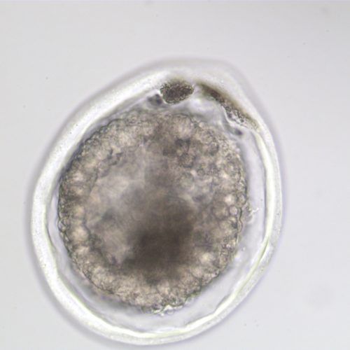
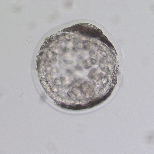
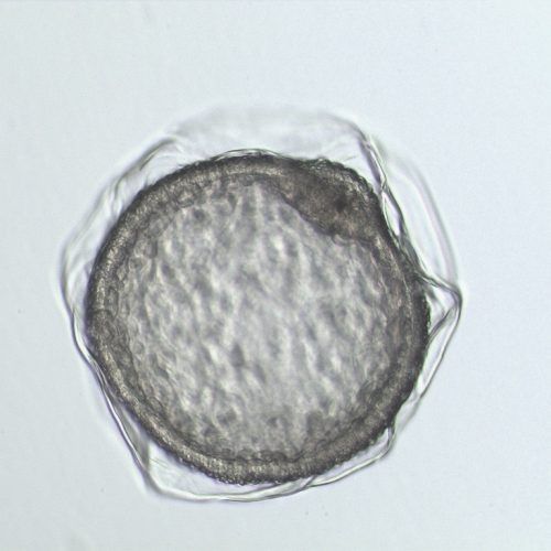
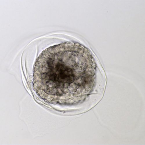
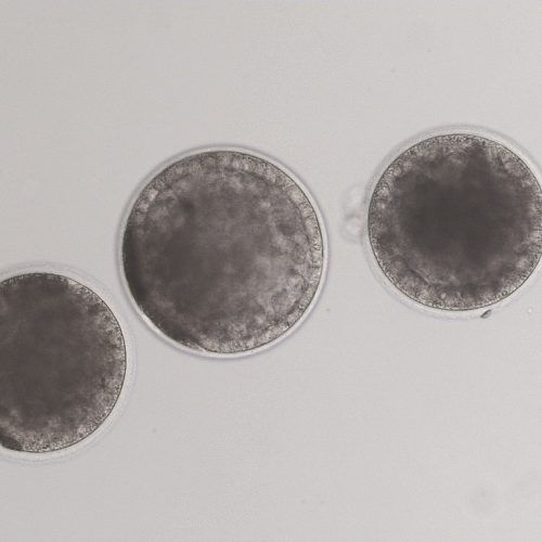
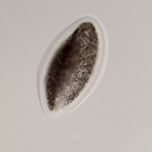
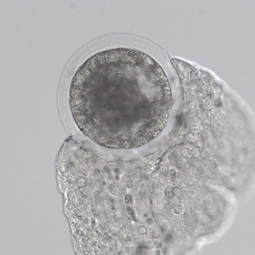
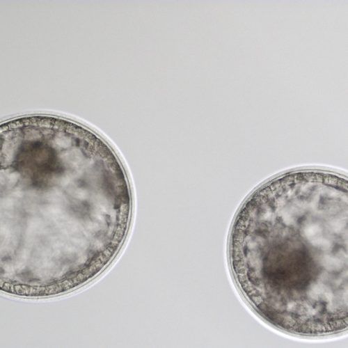
The Equine Embryo: A Gallery
The equine embryo is special in many ways:
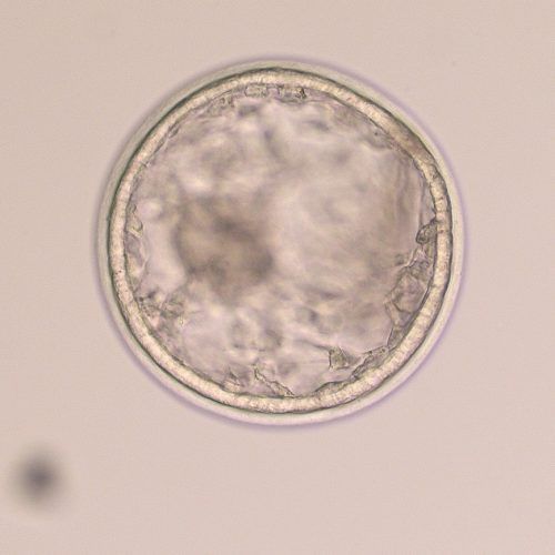
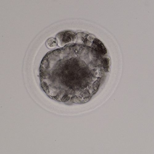
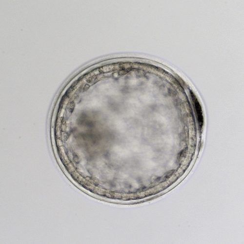
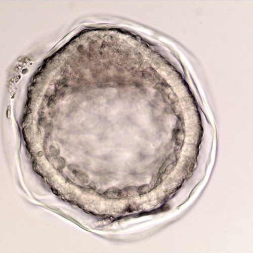
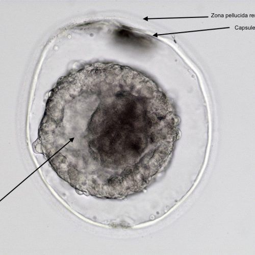
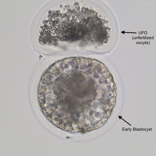
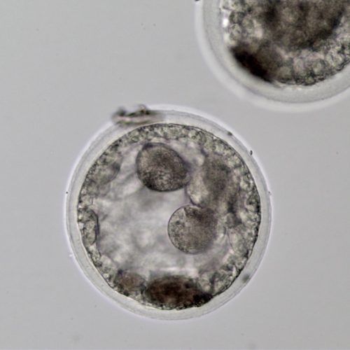
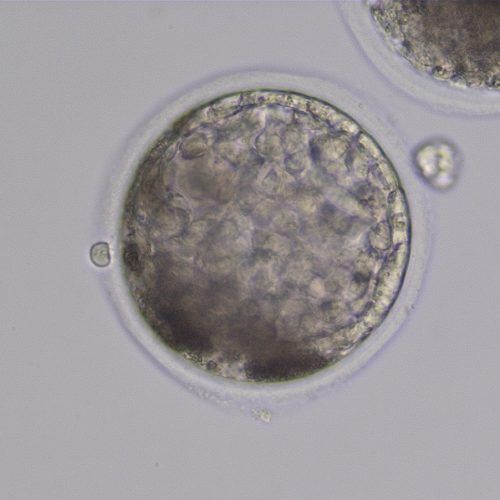
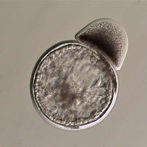
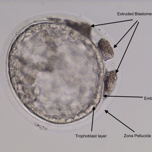
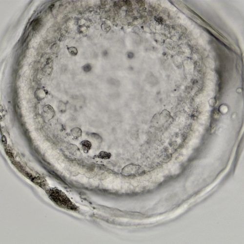
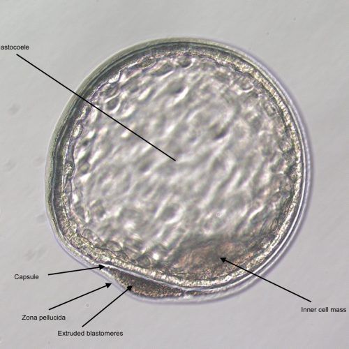
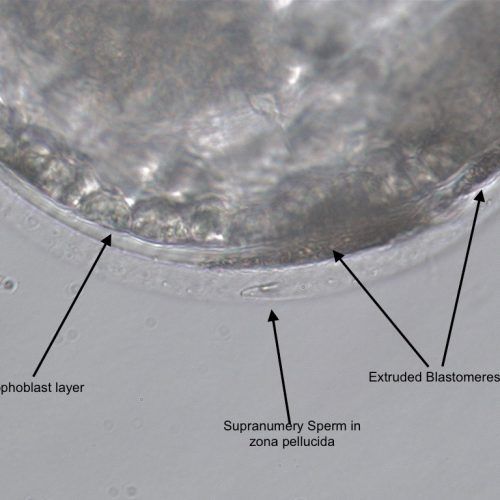
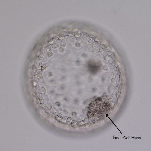
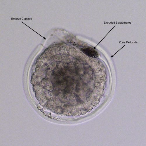
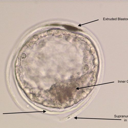
- Capsule: The embryonic capsule is unique among domestic mammals. This is a glycoprotein protective coat formed by the trophoblast layer beneath the zona pellucida. It forms only after the embryo enters the uterus, so embryos grown in a laboratory do not develop the capsule until after they are transferred. If not protected by either the zona pellucida or the embryo capsule, an embryo cannot survive in the uterus.
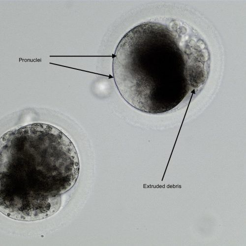
- Selective transport: Fertilization occurs and the embryo is formed within the oviduct. It remains in the oviduct for the first five and a half days after fertilization, until it signals the oviduct to move it into the uterus with the production of a specific form of prostaglandin. Without this signal, the oviduct tends to retain ova that are not fertilized or embryos that do not reach this stage of development. This means, unlike most domestic animals, unfertilized equine oocytes will generally not be recovered on a uterine flush unless they have accompanied an embryo into the uterus.
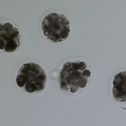
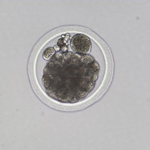
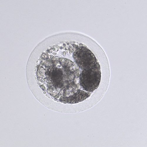
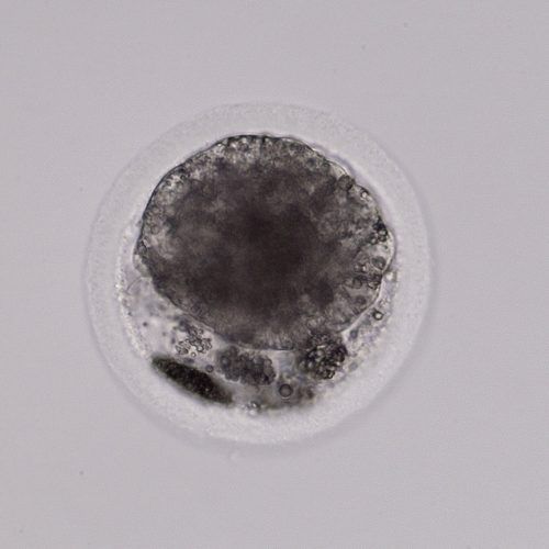
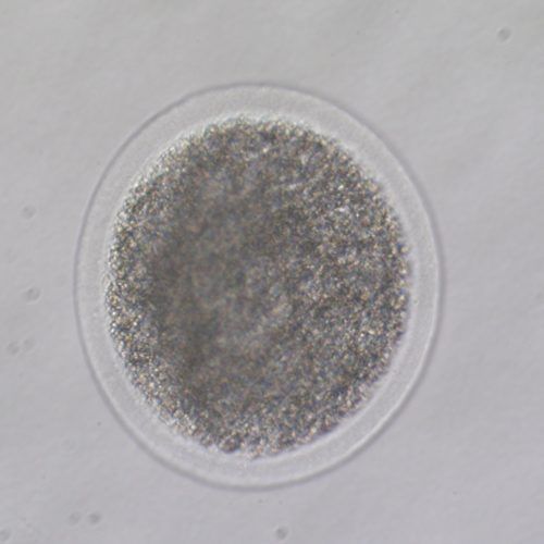
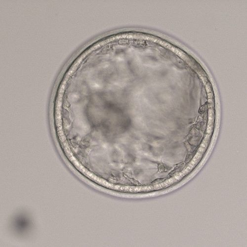
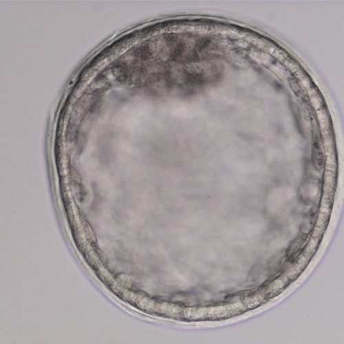
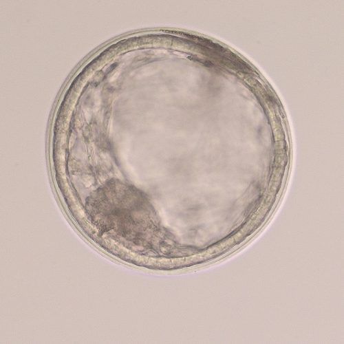
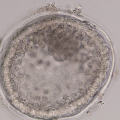
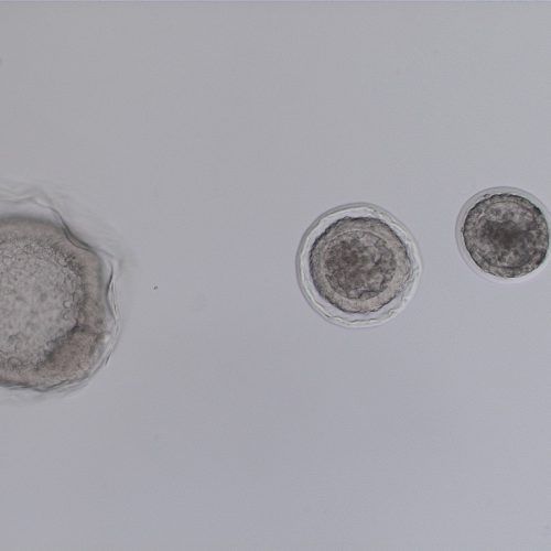
- Growth rate: Once the embryo becomes a blastocyst and starts expanding around day seven, the embryo will nearly double in size each day until day 12. This allows early ultrasound determination of pregnancy status, as an embryo around 300-400 microns (0.3-0.4mm) in size when recovered at day seven will grow to 3-4mm, large enough to be visualized on an ultrasound scan, by day 11.
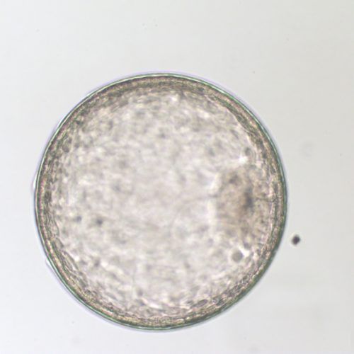
- Mobility phase: The early equine embryo does not sit quietly in the uterus, but moves rapidly along the entire endometrial surface until approximately day 17, when it becomes too large to move. The embryo needs to have close contact with the endometrial surface to stimulate maternal recognition of the pregnancy, so the mare will maintain a good environment for the embryo and not return to heat.