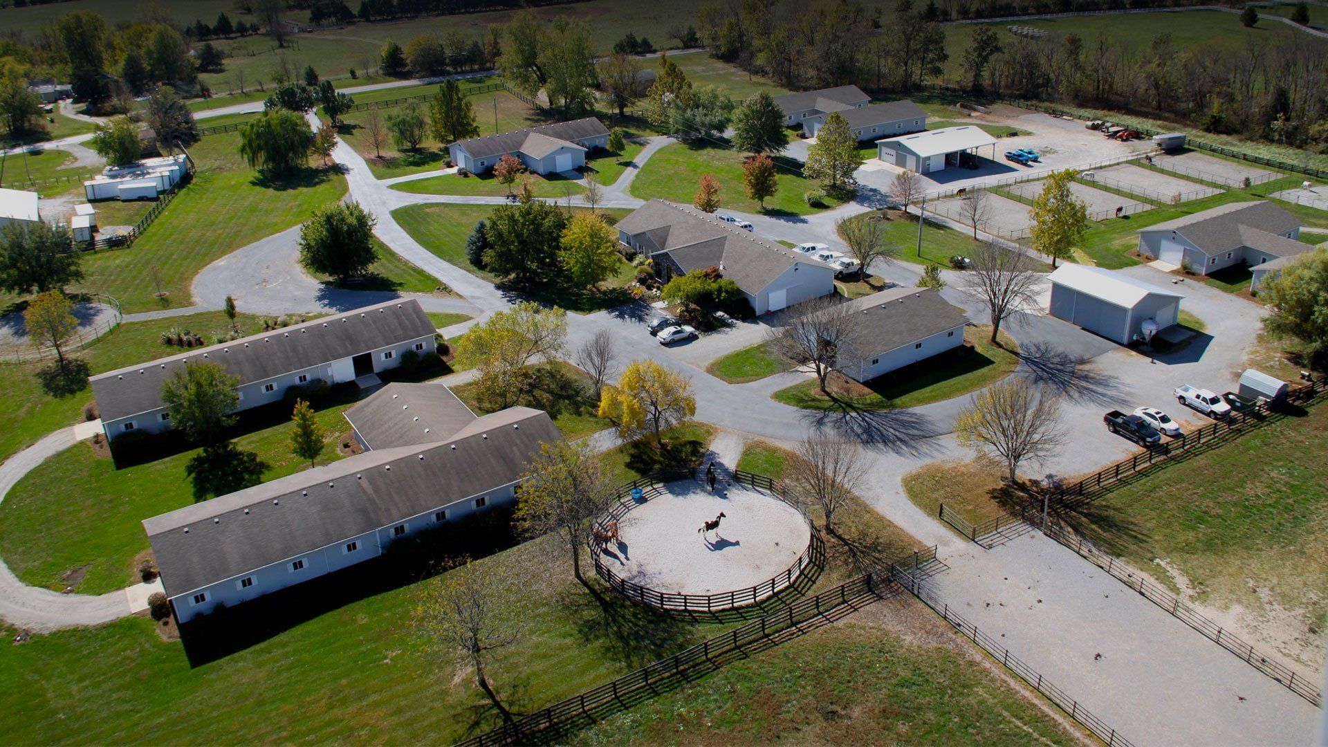
Red Maple Leaf Toxicity

August 15, 2006 by Nathan Voris, DVM
Filed under: Articles, Red Maple Leaf, Toxins 

Nathan Voris, DVM
printer friendly PDF format

Red Maple LeafAs another hot, busy summer draws to a close we enter into one of my favorite seasons-FALL. There are several reasons for my affinity for the season: mild weather, college football, and the spectacular display of fall colors offered by the many species of trees here in the Midwest. One of the most visually appealing species unfortunately is also one of potential concern to horse owners-the Red Maple.

Rest assured that not all maple trees are toxic, and not all red leafed maples are Red Maple Trees. The red maple is identified by its uniquely shaped three-pointed leaf. The exact toxic component is not known. Green leaves growing on the tree are not toxic; however, if dry, wilted leaves are eaten by a horse, the toxin causes red blood cells to break down (hemolysis).

It takes only one to three pounds of dry, wilted leaves for a 1000 pound horse to suffer the toxic effects of the red maple leaf. Clinical signs include weakness, depression, blood–tinged urine, and yellow or brown gums. Horses typically show clinical signs within 48 hours of eating the leaves. Severely affected horses die within five days. The only treatment for red maple leaf toxicity is supportive care; including intravenous fluids, blood transfusions, and oxygen therapy. Because red maple leaf toxicity compromises the horse’s ability to carry oxygen in its blood, affected horses should be kept quiet in a stall while being treated. A secondary complication of red maple leaf toxicity is acute kidney failure due to the by-products of red blood cell breakdown (methemoglobin).

Now before you cut down all of the red maple trees within a mile of your pasture, read on, there are preventative measures. Horses only actively seek tree leaves when there is nothing else to eat or they are bored. Always make sure there is adequate forage (hay or grass) in all pastures, especially those with red maples nearby. Pay close attention to your trees immediately after storms to locate fallen branches, as wilted leaves on the severed limbs are particularly toxic. If your red maple trees need pruning, it’s advisable to wait until all the leaves have fallen before cutting unwanted branches.

If you do have a tree you suspect is a red maple, you can take some leaves to your veterinarian or local nursery and landscape expert for identification. If you have a red maple tree near your horses, simply pay close attention to fallen limbs and the quality of forage available in the pasture. If you are one that will lose sleep knowing there is a red maple, have it removed and replace it with a more horse-friendly species. Whatever you decide to do with your tree, enjoy the mild weather, college football and fall colors-even those of the Red Maple.

Hyperkalemic Periodic Paralysis (HYPP)

August 7, 2006 by Hunter Ortis, DVM
Filed under: Articles, Emergency, Genetic Disorders, HYPP, Nutrition 

Hunter Ortis, DVM
printer friendly PDF format

In recent years Hyperkalemic Periodic Paralysis (HYPP), has been a familial disorder affecting many pure and crossbred quarter horses, paint horses and appaloosa horses. The disorder has been linked to the quarter horse stallion Impressive and has been genetically passed on. Impressive-bred horses were favored by many due to their heavy muscling and success in the halter show ring.

Muscle contractions are stimulated by changes in sodium and potassium levels at the cellular level. This is controlled by lots of tiny cellular pumps that move sodium and potassium in and out of the cell. During muscle contraction there is a higher level of sodium inside the cell and lower level of potassium outside the cell and for a relaxed muscle the opposite is true. In a HYPP affected horse there is a genetic defect or mutation that results in pumps that malfunction periodically causing a sustained high level of sodium in the cell and a high level of potassium outside of the cell leaving the horse with uncontrolled muscle contractions.

A blood or, more commonly now, a hair sample can be submitted to a diagnostic lab to be evaluated to see if a horse has the genetic potential to carry the disorder. Results will be reported as H/H, N/H, or N/N. Horses with H/H or N/H have the potential to have the disorder and also pass it on to their progeny. If a horse is H/H, it received the gene from both its sire and dam. An H/H horse will pass the gene on to 100% of all its foals. If a horse is N/H it received the gene from either its dam or sire and will pass the gene on to 50% of all its foals. Horses that are N/N, do not have the disorder nor do they have the genetic potential to pass the disorder on to their progeny.

The AQHA is taking steps in reducing the number of horses affected by the disorder by placing new registration restrictions regarding HYPP. According to the 2006 Official Handbook, registration rule 205(c)(3), “Effective with foals born on or after January 1, 2007, all descendants of the stallion Impressive, AQHA registration number 0767246, shall be required to be parentage verified and HYPP tested, subject to the conditions in (c)(2) above. Any foal testing homozygous positive for HYPP (H/H) will not be eligible for registration with AQHA.” Rule 205(c)(2) states that HYPP testing is not required if “the foal’s closest ancestors, tracing to the HYPP line, have been tested negative and designated on their registration certificates, these foals will automatically be designated “N/N” on their registration certificate.”

Horses that are H/H or N/H may or may not show clinical episodes of the disorder. Usually if episodes are going to occur they will begin by 3-4 years of age. Although episodes are often unpredictable there are common stimuli that may precipitate episodes such as trailer rides, stress, and fasting. Most episodes begin with a period of muscle fasciculations of the neck and head, sweating and third eyelid prolapse. There is commonly a period of muscle weakness during episodes that may progress to the horse lying down or dog sitting. Horses usually remain bright and alert and respond to stimulation. In severe episodes respiratory distress associated with “roaring sounds” may occur and even death.

All horses that have Impressive in their lineage should be tested so owners will know if there is potential for HYPP episodes. If a horse is having an episode the owner should contact their veterinarian immediately. Commonly veterinarians will give calcium gluconate intravenously at a slow rate and or dextrose intravenously to rapidly drive the potassium back into the cell. The veterinarian will also be able to evaluate the horse to see if there is other supportive care needed and give you advice on hopefully preventing further episodes such as using acetazolamide, a diuretic that helps decrease excess potassium.

If you have an H/H or N/H horse that has episodes there are a few steps you can take to help prevent episodes.

1.Avoid feeding feeds that are high in potassium such as alfalfa hay, brome hay, molasses, soybean oil and canola oil. Replace with feeds such as timothy or bermuda grass hay, grains and beet pulp.
2.Feed multiple small meals each day instead of just once or twice daily.
3.Regular exercise or being allowed access to large pasture or paddock is helpful in preventing episodes.
These steps may not control episodes in all affected horses, but astute owners, who notice early signs, can minimize an episode by administering corn syrup orally (The syrup will trigger an insulin release driving the potassium back into the cell).

Since this is a heritable trait it is recommended not to use horses that are H/H or N/H as breeding animals. In light of the recent AQHA rule changes, breeders risk having an unregisterable foal when breeding N/H to N/H or N/H to H/H. If you own a horse that has episodes work with your veterinarian to come up with a plan with treatment and prevention.

Lacerations Involving Joints

August 7, 2006 by Nathan Voris, DVM
Filed under: Articles, Emergency, Joint, Laceration, Lameness, Skin 

Nathan Voris, DVM
printer friendly PDF format

Anyone who has owned a horse knows the term “horse-safe” is an oxymoron; many horses could find a way to hurt themselves in a barn with padded stalls. In equine practice, lacerations represent one of the most frequent emergency calls. Most of the time, the wound is in a benign area that can be easily cleaned up and sutured. Other times, location of the wound combined with critical structure involvement forbids simple primary closure and calls for aggressive medical and possible surgical intervention to facilitate healing.

There are very few wounds that are as threatening to a horse’s career, or even life, than one that involves a joint. The reason for this is the anatomic and physiologic makeup of the joint itself. Beneath the skin, the joint is protected by a fibrous tissue structure called the joint capsule. On the inner side of the joint capsule is a tissue layer that produces synovial fluid. The synovial fluid is responsible for lubricating and nourishing the joint surfaces. Unfortunately, the properties that allow synovial fluid to nourish the joint also make it a perfect medium for bacterial growth if the protective layers are penetrated.

The body’s natural response to a wound is to send scavenging white blood cells to “clean up” damaged tissue and bacteria. This is the reason a joint may become swollen, hot and painful following an injury. Unfortunately, the body’s response to injury is almost as bad as the infection itself because the flood of white blood cells into a joint can lead to cartilage breakdown. The longer the infection lasts, the more likely there will be significant permanent damage to the joint.

Intra-articular irrigationLacerations near joints warrant immediate thorough evaluation by a veterinarian to assess the extent of tissue involvement. If the injury is recent and there is minimal subcutaneous inflammation (cellulitis) a needle should be inserted into the joint, on the side opposite the wound, and sterile saline should be injected to distend the joint. If saline from within the joint leaks out through the wound, the joint should be considered infected and aggressive treatment should be initiated.

Appropriate treatment involves intra-articular irrigation, broad spectrum systemic and intra-articular antibiotics, systemic anti-inflammatories, and sterile bandaging. Most of the time, the wound should not be closed with sutures due to the risk of sealing bacteria into the joint or surrounding tissues. Duration of treatment is solely dependant upon the horse’s response to treatment. If appropriate treatment is initiated within 24 hours of injury, horses have an 85% chance of survival and 50% chance of returning to full function.

Sometimes a cut that appears to be “a long way from the heart” can be a big problem but rapid identification of joint involvement and aggressive medical and surgical care can tip the odds in your favor for a positive outcome.